Some say that the eyes are the windows to the soul. Is this true? Well, I like to believe so. What I do know for certain is the eyes are a window to your overall health. Did you know that your eye doctor can see diabetes and high cholesterol, oftentimes even before your primary care doctor does?
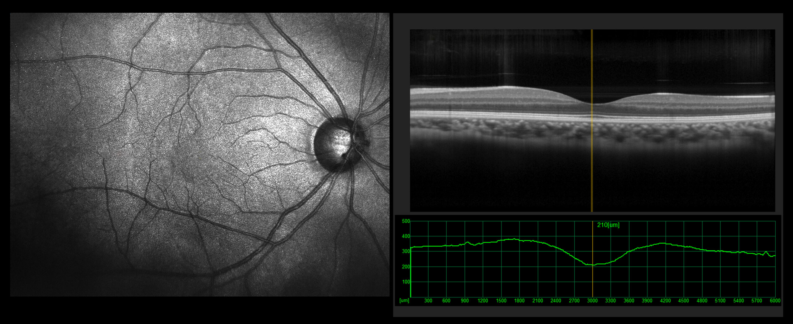
In addition to a dilated fundus exam, Thomas Eye Center offers additional screenings in order to have a more in-depth look at the health of your eyes, and your overall health. Our office recommends OCT (Optical Coherence Tomography) screenings and fundus photography for all patients.
The Power of Advanced Imaging in Optometry: Why OCT and Fundus Photography Matter
In the ever-evolving field of optometry, technology plays an increasingly vital role in diagnosing and managing eye diseases. Two non-invasive imaging techniques, Optical Coherence Tomography (OCT) and fundus photography, have revolutionized how eye care professionals assess ocular health. These advanced tools provide unparalleled insights into the structure and function of the eye, enabling early detection of sight-threatening conditions and personalized treatment plans. These also allow doctors to have a detailed comparison of the eyes from year to year and quickly see any potentially serious changes.
OCT: Seeing Beneath the Surface
Conventional eye exams are limited to what can be visually observed on the eye’s surface. OCT, however, uses low-coherence interferometry to capture high-resolution, cross-sectional images of the retina and optic nerve. This is similar to ultrasound, but uses light waves instead of sound.
These detailed scans reveal the intricate layers of the retina and optic nerve head, allowing for the detection of subtle changes that may indicate disease. OCT is invaluable in diagnosing and monitoring conditions such as age-related macular degeneration, diabetic retinopathy, glaucoma, and macular holes. By tracking changes over time, OCT guides treatment decisions and helps assess therapeutic response.
Fundus Photography: A Window to the Eye
Fundus photography involves capturing high-resolution images of the posterior segment of the eye, including the retina, optic disc, and blood vessels. These photos provide a permanent visual record of the eye’s health, facilitating longitudinal comparisons and early detection of abnormalities.
Unlike OCT, fundus photography does not produce cross-sectional images, but it offers a wider field of view, allowing for the assessment of peripheral retinal health. This is particularly useful in diagnosing conditions like diabetic retinopathy and retinal detachments. The photos can also highlight signs of systemic diseases, such as hypertension, diabetes, and cardiovascular disease, that may manifest in the eye.
The Power of Combination
While both OCT and fundus photography are powerful diagnostic tools, their true potential is unleashed when used in combination. This multimodal imaging approach provides a comprehensive understanding of both the eye’s structure and function. OCT’s cross-sectional analysis complements the widefield imaging of fundus photography, offering a more complete picture of ocular health than either modality could provide alone.
